Osteoporosis Self-Assessment Quiz - Test Your Bone Health Risk Online
Currently, osteoporosis affects over 200 million people worldwide and is responsible for more than 8.9 million fractures annually. As many as half of all women and a quarter of men over the age of 50 will suffer a fracture due to osteoporosis in their lifetime. Yet the vast majority of people with osteoporosis have no symptoms until a fracture occurs — earning it the name the "silent disease." Because bone loss occurs gradually and without pain, most people don't know they are at risk until it is too late to prevent the first fracture.
Take the Osteoporosis Self-Assessment Quiz Now!
Osteoporosis Self-Assessment | HRT.org
Osteoporosis Self-Assessment
Osteoporosis affects over 200 million people worldwide. Most people have no symptoms until a fracture occurs — making early risk assessment essential.
Answer 13 questions about your lifestyle, health history, and risk factors to understand your personal risk profile and what you can do to protect your bone health.
1
Question text…
Select the answer that applies to you.
press Enter ↵
14
What is your first name?
Please enter your first name
press Enter ↵
15
We'll calculate your results in a second — if you'd like to receive a copy via email, leave your email below:
Please enter a valid email address
press Enter ↵
16
Would you like to be notified of new assessments and health-related posts?
Osteoporosis is a skeletal disease characterised by reduced bone mineral density (BMD) and deterioration of bone microarchitecture, leading to increased bone fragility and susceptibility to fracture. The word comes from the Latin osteon (bone) and poros (passage) — literally "porous bone." Bone is a living tissue that is continuously broken down and rebuilt throughout life. Osteoporosis develops when the rate of bone resorption exceeds the rate of bone formation, resulting in a net loss of bone mass over time. The related condition osteopenia describes bone density that is lower than normal but not yet in the osteoporotic range — and without intervention, it commonly progresses to osteoporosis.
What does bone density mean?
Bone mineral density (BMD) is a measure of the amount of mineral — primarily calcium and phosphate — packed into a given area of bone. It is measured using a DEXA (dual-energy X-ray absorptiometry) scan and expressed as a T-score — the number of standard deviations a patient's BMD falls above or below the average for a healthy young adult of the same sex. A T-score of -1.0 or above is considered normal. A T-score between -1.0 and -2.5 indicates osteopenia. A T-score of -2.5 or below indicates osteoporosis. Each standard deviation reduction in T-score approximately doubles fracture risk.
What risk factors may indicate osteoporosis?
- Sedentary lifestyle or lack of weight-bearing exercise - Regularly drinking two or more alcoholic drinks per day - Current or past smoking - High caffeine intake (more than three cups per day) - Long-term use of steroids, anticonvulsants, or thyroid medication - Failure to meet daily calcium requirements through diet - Abnormal absence of menstrual periods (amenorrhoea) - Postmenopausal status, including early or surgically induced menopause - Low testosterone (in men) - Caucasian or Asian ethnic background - Family history of osteoporosis or fragility fracture - Forward curvature of the upper back (kyphosis)
Can I test my bone density online at home?
While a definitive bone density measurement requires a DEXA scan — available at most hospitals and private clinics — you can begin the assessment process at home. A bone health blood panel measuring vitamin D, calcium, parathyroid hormone (PTH), and bone turnover markers provides important information about your bone metabolism and nutritional status. You can also use the risk assessment quiz on this page to identify whether you have multiple risk factors that warrant further investigation.
Types of Osteoporosis
- Primary osteoporosis Type I (postmenopausal) — The most common form, occurring in women in the years following menopause due to the rapid decline in oestrogen. Primarily affects trabecular (spongy) bone, particularly in the vertebrae and wrist. - Primary osteoporosis Type II (age-related/senile) — Affects both men and women over age 70 due to the cumulative effects of ageing on bone remodelling, calcium absorption, and vitamin D metabolism. Affects both trabecular and cortical bone, particularly the hip. - Secondary osteoporosis — Caused by an identifiable underlying condition or medication. Common causes include glucocorticoid (steroid) use, hyperthyroidism, hypogonadism, malabsorption conditions, chronic kidney disease, and certain medications. - Osteopenia — Below-normal but not yet osteoporotic bone density. A significant fracture risk factor that should be taken seriously and managed proactively.
What osteoporosis tests can I take?
The following tests are available to assess bone health and identify contributing factors:
- DEXA scan — gold standard for measuring bone mineral density (T-score) - Vitamin D (25-hydroxyvitamin D) — deficiency is one of the most common drivers of bone loss - Calcium (serum) — to assess calcium status and parathyroid function - PTH (Parathyroid Hormone) — elevated PTH drives bone resorption; important for excluding hyperparathyroidism - Bone turnover markers — P1NP (formation) and CTX (resorption) reflect the rate of bone remodelling - Oestradiol (E2) — low oestrogen is the primary hormonal driver of postmenopausal bone loss - Testosterone (total and free) — low testosterone in men is a significant and under-recognised cause of osteoporosis - FSH and LH — to confirm postmenopausal or hypogonadal status - TSH, Free T3, Free T4 — hyperthyroidism and excessive thyroid hormone replacement both reduce bone density - Cortisol — elevated cortisol (from chronic stress or exogenous steroids) directly suppresses bone formation
Osteoporosis Tests Defined
DEXA Scan—This is the gold standard for measuring bone mineral density. It produces a T-score which compares your bone density to that of a healthy young adult of the same sex. It is a low-radiation, painless scan taking 10–20 minutes and is available at most hospitals and private scanning centres.
Vitamin D Test—This test measures 25-hydroxyvitamin D, the storage form of vitamin D. Deficiency (below 50 nmol/L) is extremely common and is one of the most easily correctable contributors to bone loss. Supplementation with vitamin D3 combined with vitamin K2 is the standard recommendation.
PTH Test—This test measures parathyroid hormone. Elevated PTH drives bone resorption and is seen in hyperparathyroidism, vitamin D deficiency, and chronic kidney disease. Identifying and treating elevated PTH can significantly reduce ongoing bone loss.
Bone Turnover Markers—P1NP measures the rate of new bone formation. CTX measures the rate of bone resorption. Elevated CTX alongside low or normal P1NP indicates accelerated net bone loss, prompting further investigation and treatment.
What Test Results Mean
DEXA T-Score—A T-score of -2.5 or below confirms osteoporosis and typically triggers treatment. Scores between -1.0 and -2.5 (osteopenia) warrant lifestyle intervention, calcium and vitamin D optimisation, and monitoring. The FRAX tool can estimate your 10-year fracture probability using clinical risk factors with or without a BMD result.
Vitamin D Test—Optimal vitamin D levels above 75 nmol/L (30 ng/mL) are essential for calcium absorption and bone mineralisation. Supplementation with vitamin D3 (1000–4000 IU/day depending on baseline levels) combined with vitamin K2 is the standard recommendation.
PTH Test—Elevated PTH drives bone resorption. Identifying and treating the cause — whether vitamin D deficiency, hypercalciuria, or hyperparathyroidism — can significantly slow bone loss.
Oestradiol and Testosterone—Low oestrogen in postmenopausal women and low testosterone in men are the leading hormonal drivers of osteoporosis. You can also assess this using our Male Hormone Home Test Kit or Comprehensive Female Hormone Home Test Kit.
Common Causes of Osteoporosis
- Postmenopausal oestrogen deficiency — the most common cause in women - Age-related bone loss (both sexes) - Low testosterone in men (hypogonadism) - Vitamin D deficiency and reduced calcium absorption - Long-term corticosteroid use (glucocorticoid-induced osteoporosis) - Hyperthyroidism or excessive thyroid hormone replacement - Hyperparathyroidism - Malabsorption conditions (coeliac disease, Crohn's disease, bariatric surgery) - Amenorrhoea (including exercise-induced, anorexia-related, or hypothalamic) - Sedentary lifestyle and lack of weight-bearing exercise - Smoking and excess alcohol consumption - Genetics and family history - Chronic kidney or liver disease
Treatment Options for Osteoporosis
Treatment for osteoporosis may include:
- Bisphosphonates (alendronate, risedronate, zoledronic acid) — reduce bone resorption and fracture risk - Denosumab (Prolia) — RANK ligand inhibitor that reduces osteoclast activity - Hormone replacement therapy (HRT) — particularly effective for postmenopausal women; reduces bone loss and fracture risk with decades of supporting evidence - Testosterone replacement therapy (TRT) — for men with confirmed hypogonadism and low bone density - Teriparatide (PTH analogue) — anabolic therapy that stimulates new bone formation; used in severe or refractory osteoporosis - Romosozumab — sclerostin inhibitor; both anti-resorptive and bone-building - Calcium and vitamin D3 supplementation — foundational for all patients; dosing guided by baseline levels - Vitamin K2 (MK-7) — directs calcium into bone and away from arterial walls
Natural support may also include regular weight-bearing and resistance exercise (the single most effective lifestyle intervention for maintaining bone density), a calcium-rich diet (dairy, leafy greens, sardines), adequate vitamin D through sunlight and supplementation, reducing alcohol and caffeine intake, and smoking cessation. Reducing fall risk through balance training, home safety modifications, and vision correction is as important as pharmacological treatment for preventing fracture-related morbidity.
Based on your osteoporosis assessment and risk factors, further investigation including a DEXA scan and hormone panel may be indicated.
See a healthcare practitioner to discuss your risk factors and possible testing, or order tests on your own and share results with your doctor.
References
1"Osteoporosis prevention, diagnosis, and therapy." NIH Consensus Development Panel on Osteoporosis. JAMA. 2001;285(6):785–795. 2"Management of osteoporosis in postmenopausal women: the 2021 position statement of The Menopause Society." Menopause. 2021;28(9):973–997. 3Kanis JA, et al. "European guidance for the diagnosis and management of osteoporosis in postmenopausal women." Osteoporos Int. 2019;30(1):3–44.
A deficiency of bone-protective hormones — particularly oestrogen, testosterone, and vitamin D — can affect virtually all aspects of skeletal health. The severity of bone loss ranges from mild osteopenia to severe osteoporosis with multiple fragility fractures. It is estimated that 1 in 2 women and 1 in 4 men over 50 will experience an osteoporotic fracture in their lifetime, many of whom were unaware of their condition.
Self-assessment Questionnaire
Because osteoporosis produces no symptoms in its early stages, risk factor assessment is the primary tool for identifying people who need further investigation. The risk factors assessed in the questionnaire above — including exercise habits, dietary calcium, hormonal status, smoking, alcohol, medication use, and family history — are the same factors used in validated clinical risk assessment tools. Multiple risk factors substantially increase fracture probability and should prompt bone density testing.
We strongly suggest that you contact your healthcare provider if you have any concerns about your bone health or risk of osteoporosis. The self-assessment above should not be substituted for bone density testing by your provider.
FAQs about Osteoporosis Assessment Quiz and Online Tests
How do you perform an osteoporosis assessment?
An osteoporosis assessment combines a clinical risk factor evaluation — such as the questionnaire on this page — with a DEXA scan for definitive bone mineral density measurement. Blood tests measuring vitamin D, calcium, PTH, and bone turnover markers help identify the biochemical drivers of bone loss. The FRAX tool can estimate your 10-year fracture probability using clinical risk factors with or without a BMD result.
Are home bone health tests accurate?
Yes, home blood tests for bone health markers — including vitamin D, calcium, PTH, and hormone levels — are accurate when processed by certified laboratories. They are a valuable and accessible first step in understanding your bone health status and the hormonal and nutritional factors driving bone loss. A DEXA scan remains the gold standard for definitive bone density measurement.
Do online osteoporosis tests work?
Online osteoporosis risk assessments and at-home bone health blood panels are effective screening tools. Validated risk assessment questionnaires have good sensitivity for identifying people with low bone density and are used clinically to decide who should proceed to DEXA scanning. A comprehensive blood panel identifies the underlying hormonal and nutritional drivers that a DEXA scan alone cannot reveal.
Are there any physical signs of osteoporosis?
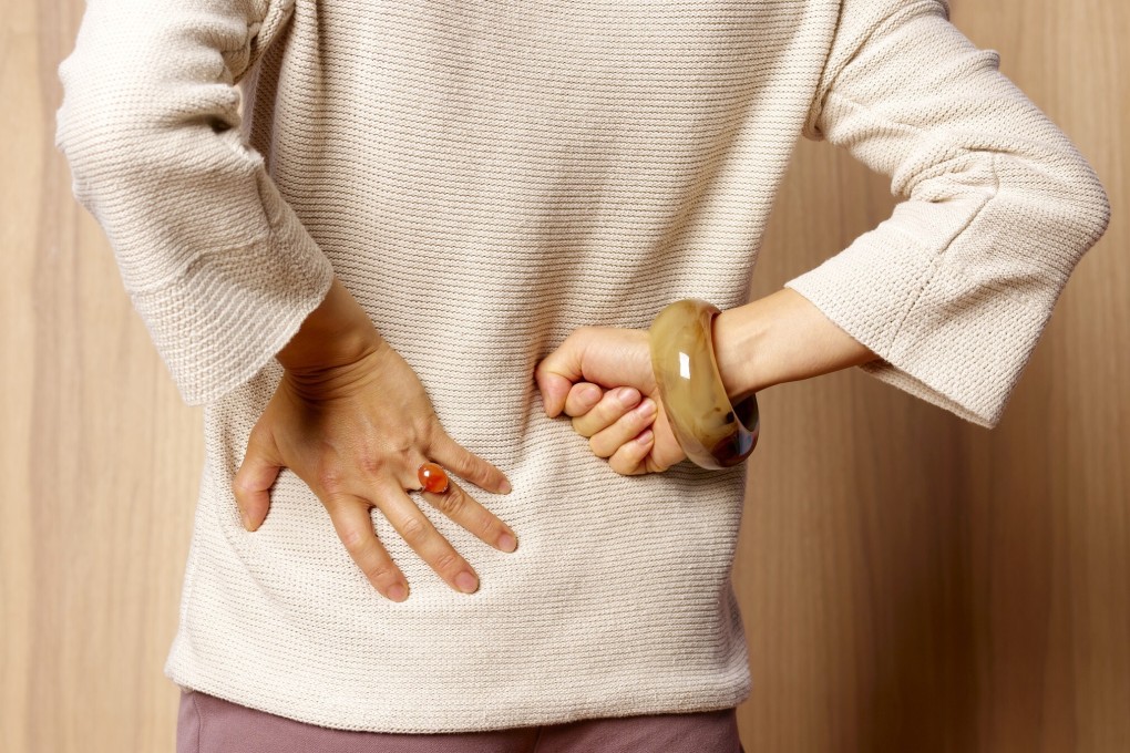
Yes, while osteoporosis typically produces no symptoms until a fracture occurs, some later-stage signs include loss of height (more than 2cm over time), forward curvature of the upper back (kyphosis or "dowager's hump"), and back pain from vertebral compression fractures. Fragility fractures — fractures that occur from a fall from standing height or less — are the most common first clinical presentation. The wrist, spine, and hip are the most commonly affected sites.